Examinations in angiology (vascular medicine)
- Vascular ultrasound (Doppler/duplex)
- Ozillography
- ABI measurment
- Early detection of arteriosclerosis with PWV
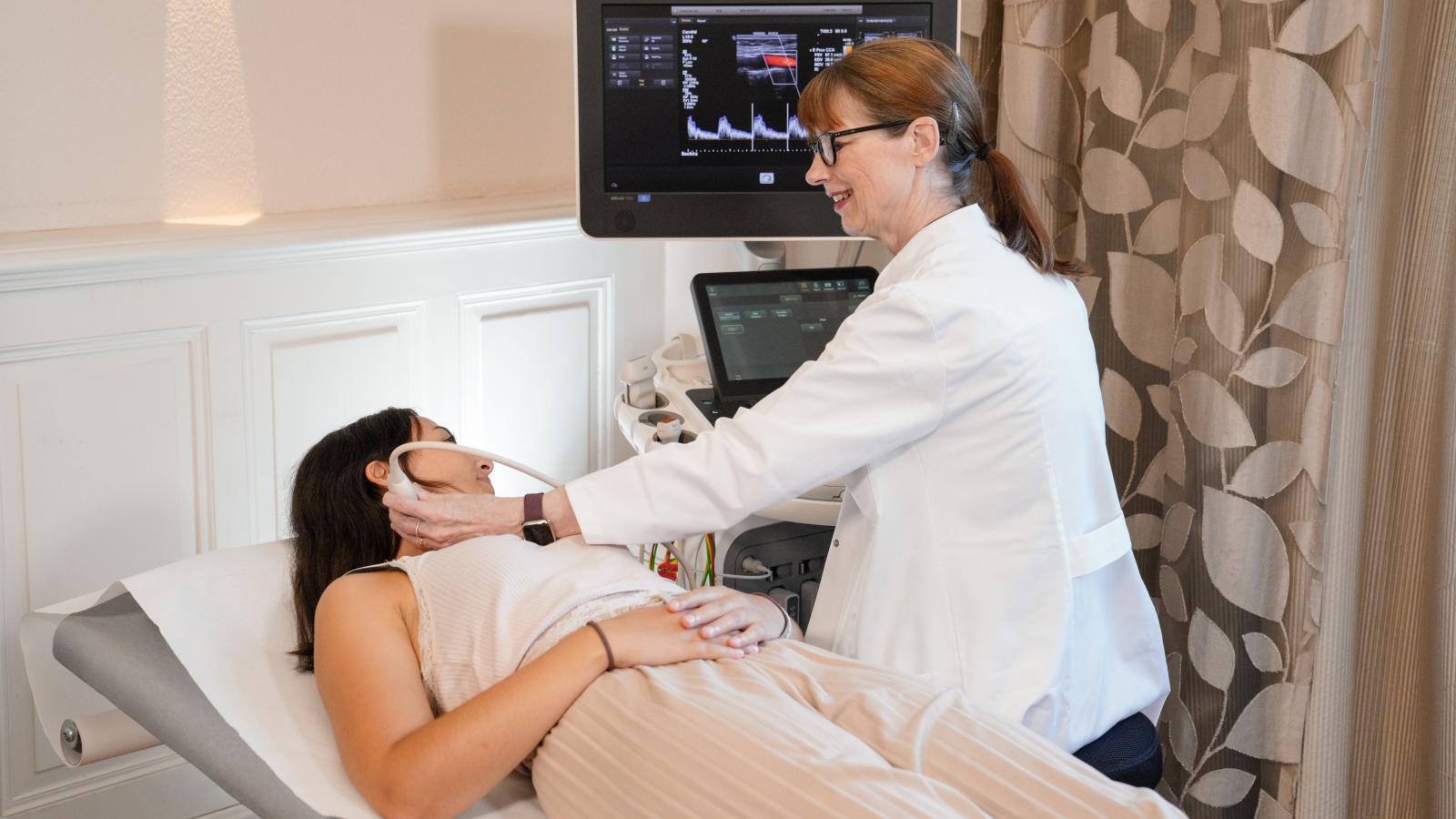
Vascular ultrasound (Doppler/duplex)

Ultrasound is an elementary diagnostic tool in angiology. The examination allows the vessels to be visualised non-invasively (in contrast to angiographic vascular imaging using X-rays with catheter procedures). The advantages are: no X-rays are used, no contrast medium harmful to the kidneys is required and the examination is absolutely painless.
The examination serves to assess, among other things:
- Calcifications, constrictions (stenoses), vascular occlusions and thus circulatory disorders in arteries
- Vascular dilatations (aneurysms)
Oscillography
If a circulatory disorder of the leg arteries is suspected, segmental oscillography is performed in addition to the ABI measurement. The measurement is comparable to a blood pressure measurement. Cuffs are applied to the upper and lower legs as well as the forefoot, then the blood flow volume is measured and visualised using measurement curves. Depending on the symptoms, oscillography can also be performed on the fingers or toes. This detailed analysis enables the exact localisation of constrictions or occlusions in the vessels. An ultrasound examination is then carried out in the affected vascular area to look for the narrowing and determine the appropriate treatment.
ABI measurement
The ABI is the ankle-brachial index. Similar to blood pressure measurement, cuffs are applied to both the wrists and ankles in order to simultaneously record the volume fluctuations in these areas during the cardiac cycle. This measurement can be supplemented with various stresses (knee bends, toe stands) in order to assess the blood flow under the influence of muscular activities. The resulting ankle-brachial index serves as a screening test for the presence of peripheral arterial occlusive disease (PAD) and also has prognostic significance for cardiovascular events such as heart attack and stroke.
Early detection of arteriosclerosis with PWV
The pulse wave velocity (PWV) is the speed at which the pressure wave travels through the arteries of an organism. The clinical picture of arteriosclerosis is one of premature vascular ageing. The deposition of substances on the blood vessels leads to increased vascular stiffness, which can be recognised by an increased PWV.
The examination is painless. As with the ABI measurement, cuffs are placed on both wrists and ankles, and an ECG clamp is also attached to each upper arm. The pulse wave velocity (PWV) can provide information about the mortality rate in diseases such as diabetes mellitus or terminal renal failure due to the changes in the vascular system associated with pathological values. It can also help to assess general cardiovascular risk factors.